

an email newsletter released every month highlighting the latest articles, events, technical inquiries, and voices from the community
Technology Readiness Level (TRL) 6 Device to Identify Traumatic Brain Injury (TBI)

https://www.dvidshub.net/search?q=TBI&view=grid
Posted on October 24, 2019 | Completed on October 24, 2019
What are the technology readiness level (TRL) 6 devices that could aid in the diagnosis of traumatic brain injury (TBI)?
1. Inquiry
HDIAC received the following technical inquiry from United States Army Materiel Systems Analysis Activity (AMSAA):
“We are looking for TRL 6 Prototype devices that could aid in the diagnosis of TBI far forward on the battlefield, including the ability to assess the existence and severity of brain trauma, i.e., intracranial bleeding.”
2. HDIAC Response
TBI usually results from a violent blow or jolt to the head or body. An object that penetrates brain tissue, such as a bullet or shattered piece of skull, also can cause TBI. Mild TBI may affect brain cells temporarily. However, more serious TBI can result in bruising, torn tissues, bleeding, and other physical damage to the brain. Explosive blasts are a common cause of TBI in active-duty military personnel. Although how the damage occurs isn’t yet well understood, many researchers believe that the pressure wave passing through the brain significantly disrupts brain function. TBI also results from penetrating wounds, severe blows to the head with shrapnel or debris, and falls or bodily collisions with objects following a blast.
TBI poses a grave threat to the ability of servicemen and women to function on the battlefield, as well as hinders their ability to achieve full potential later in life. The “golden hours,” or the treatment administered during the initial hours after injury, plus the 24 to 48 hours of care afterward, are critical, as they can determine whether a patient survives their injury and, if so, what type of long-term function and disability issues they may have. As such, rapid diagnosis to determine if a brain injury has or may have occurred, forward on the battlefield, is critical to ensure appropriate treatment in those initial “golden hours.” During this research, several devices were identified to aid in this diagnostic process; additionally, individuals who are working on the development of prototypes are also identified.
3. Equipment With FDA Approval Licenses
3.1 Infrascanner Model 2000
Description: A portable screening device that uses near-infrared (NIR) technology to screen patients for intracranial bleeding, identifying those who would most benefit from immediate referral to a CT scan and/or neurosurgical intervention. It is likely beyond TRL 6, as it has been tested and utilized by the U.S. Marine Corps in its battalion aid stations; however, it is added here, as it provides a simple graphic report that gives a positive or negative indication of concussion or brain bleed.
Accuracy: In patients with epidural, subdural, and intracerebral hematomas: sensitivity = 88% / specificity = 90.7% [1].
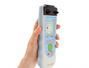
Figure 1: Infrascanner Model 2000
3.2 Banyan Biomarkers’ Brian Trauma Indicator, or BTITM
Description: The BTI can identify two brain-specific protein markers, called UCH-L1 (Ubiquitin Carboxy-terminal Hydrolase-L1) and GFAP (Glial Fibrillary Acidic Protein). These proteins rapidly appear in the blood and are elevated 12 hours following an incident where a head injury occurs and can signify if there is bleeding in the brain. The new blood test to detect the proteins will be based on Philips’ Minicare I-20 system.
The system consists of a handheld analyzer, dedicated software, and a single-use, disposable cartridge containing the application-specific test. Based on Philips’ proprietary Magnotech biosensor technology, the Minicare I-20 system is being developed to detect multiple target molecules at low concentrations with a blood sample and to show the results on the analyzer display within minutes [2].

Figure 2: BTI used with Minicare I-20
3.3 BrainScope Ahead 100, 200, 300, 400, One, Modified One
Description: BrainScope® is a medical neurotechnology company that is pioneering the assessment of brain injury, including concussion. The company’s innovative BrainScope One system is an easy-to-use, non-invasive, hand-held platform that empowers physicians to quickly make accurate head injury assessments at the point of care.
BrainScope’s unique system leverages advanced digital signal processing, sophisticated algorithms, and machine-learning technologies to identify and evaluate key brain electrical activity biomarkers of TBI. BrainScope One also includes digitized versions of standard clinical assessments for milder forms of TBI known as concussion [3].

Figure 3: BrainScope One
3.4 SyncThink in Partnership With the Brain Trauma Foundation
Description: An assessment battery to record and analyze eye-tracking impairment, EYE-SYNC® is backed by 15 years of clinical research and features lightweight, portable, fast, and objective results. The EYE-SYNC platform is a modified VR headset with infrared cameras that connect wirelessly and securely to a tablet where results are viewable to the clinician within 60 seconds.
The comprehensive brain health platform includes EYE-SYNC Smooth Pursuit, EYE-SYNC VOR, and EYE-SYNC Saccade-based assessments. Through these assessments and based on extensive peer-reviewed research, abnormal patterns can be detected that are indicative of a particular visual impairment in need of targeted treatment intervention [4].

Figure 4: Eye-SYNC
4. Ongoing Research
4.1 Trans Ocular Brain Impendence (TOBI) Prototype
Description: The TOBI Prototype is under development by Dr. Tiba and his team at the University of Michigan Center for Integrative Research in Critical Care. Uses electrical bioimpedance and ultrasound in a novel manner that will not only evaluate intracranial pressure and cerebral blood flow but also monitor cerebrovascular autoregulation (CAR), one of the most important neuroprotective processes of the brain. They were awarded funding through the “Massey TBI Grand Challenge Family Foundation Competition,” which provides funding to a select group of researchers on high-risk critical care.
This technology is non-invasive, does not require an experienced operator, and is compact in size, meaning it can be used by caregivers across all echelons of care. TOBI could provide valuable early information on CAR to help inform the diagnosis and treatment of TBI. It may be of interest that the Joyce Massey TBI Summit at the University of Michigan annually hosts scientists to present cutting-edge TBI research and exchange ideas [5].
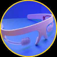
Figure 5: TOBI Prototype
5. Wearable Monitors
5.1 NoMo Cyton-Based EEG Headset Prototype
Description: A headset developed by NoMo Diagnostics associated with Columbia University. What sets the NoMo device apart from previous accelerometer-based detection helmets is that it monitors the brain’s actual physiological activity and change in electromagnetic signal in response to these hits that may cause TBI [6].
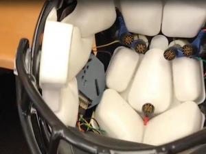
Figure 6: NoMO EEG Headset Prototype
5.2 BlastGauge
Description: Under a DARPA contract, the Rochester Institute of Technology (RIT) developed the BlastGauge, a small device worn by Warfighters to measure blast exposure and cue medics for initial response. It is a small, self-contained system that quantifies the amount of blast exposure to which a Warfighter has been exposed by indication of green, yellow, or red light [7].
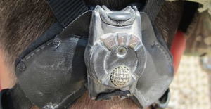
Table 7: BlastGauge
5.3 Fort Bragg Defense Veterans Brain Injury Center Wearable Sensor
Description: A current DVBIC project for development of a wearable sensor for rapid assessment of acute mild TBI based on cardiac response. The goal of the project is to assess the efficacy and sensitivity of a field-deployable system that uses cardiac measures to identify mild traumatic brain injury (mTBI). [8]
6. Rapid Testing Device
6.1 Point of Care Test (POCT) TBIcheck
Description: Dr. Sanchez of Geneva University is analyzing protein levels in the blood to possibly diagnose with a single drop of blood the possibility of an mTBI. Four molecules indicate the presence of a brain injury: H-FABP, Interleukin-10, S100B, and GFAP. If a lane appears, the injured person must go to a hospital for a CT scan; if there is nothing, the person can go home safely.
The device involves a rapid diagnostic test (POCT) called TBIcheck, inspired by the principle of pregnancy testing – by placing a single drop of blood on the well of a small 5-cm plastic case, the patient knows within 10 minutes whether there is a risk of mild trauma, namely whether or not his H-FABP level is higher than 2.5 nanograms per milliliter of blood [9].
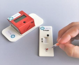
Figure 8: TBIcheck
7. Related Research of Note
During this investigation, research regarding pre-emptive brain protection was discovered, as well as a recent proposal for a TBI portable detection device from researchers at the University of Michigan. These are included here as they may be of interest to AMSAA.
7.1 Intranasal Insulin Therapy to Protect the Brain
Description: Dr. Neumar and Dr. Tulasi Jinka of the University of Michigan are developing intranasal insulin after TBI. Intranasal insulin, or insulin nasal spray, is already in clinical trials for Alzheimer’s disease and stroke, but researchers hope to prove it can successfully protect the brain when a high dose is given to a TBI patient soon after injury [10].
• The nasal spray speeds up the ability for the hormone to get to the brain through small blood vessels in the nose instead of waiting for it to enter and go through the bloodstream when administered in the arm or stomach.
• The research team will study the neuroprotective effects of an early dose and a treatment strategy where non-medical providers could administer the dose to a TBI patient right after an injury takes place.
7.2 Point-of-Care Device to Monitor and Measure Biomarkers in Real-Time
Description: Dr. Mark Burns and Dr. Frederick Korley at the University of Michigan proposed a portable, point-of-care device that could measure biomarkers in the fluid and blood and display the results on a handheld device, such as a smartphone, within 15 minutes.
• A microfluidic device for performing repeated measurements of identified blood-based biomarkers at the point of care is being developed to examine cerebrospinal fluid. Blood contains biomarkers that researchers say could play a role in how a TBI is diagnosed and monitored during the progression of the injury [10].
8. References
[1] https://infrascanner.com/models/
[2] https://www.philips.com/a-w/about/news/archive/standard/news/press/2016/20160107-Philips-and-Banyan-Biomarkers-partner-to-develop-and-commercialize-new-handheld-blood-test.html
[3] https://brainscope.com/products
[4] https://syncthink.com/
[5] https://mcircc.umich.edu/ocular-impedance
[6] https://openbci.com/community/cyton-based-eeg-headset-prototype-used-to-detect-brain-injury-in-real-time/
[7] https://www.darpa.mil/news-events/2012-05-21
[8] https://dvbic.dcoe.mil/research/wearable-sensor-rapid-assessment-acute-mild-tbi-based-cardiac-response/
[9] https://www.techexplorist.com/new-portable-device-capable-diagnosing-mild-traumatic-brain-injury-10-minutes/15803/
[10] https://labblog.uofmhealth.org/health-tech/research-into-innovative-treatment-ideas-for-traumatic-brain-injury/
Want to find out more about this topic?
Request a FREE Technical Inquiry!